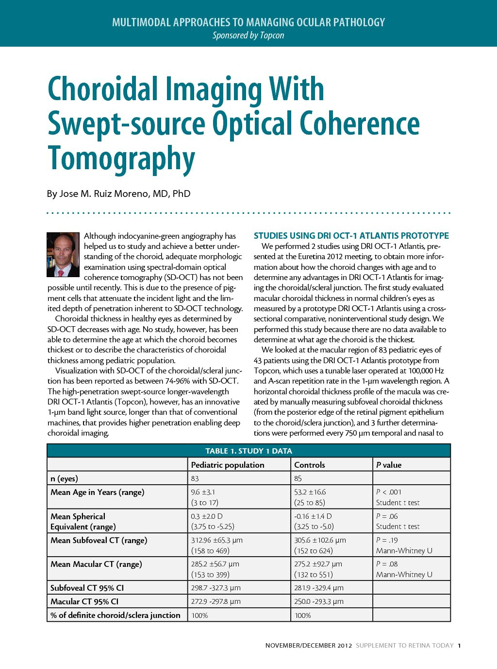
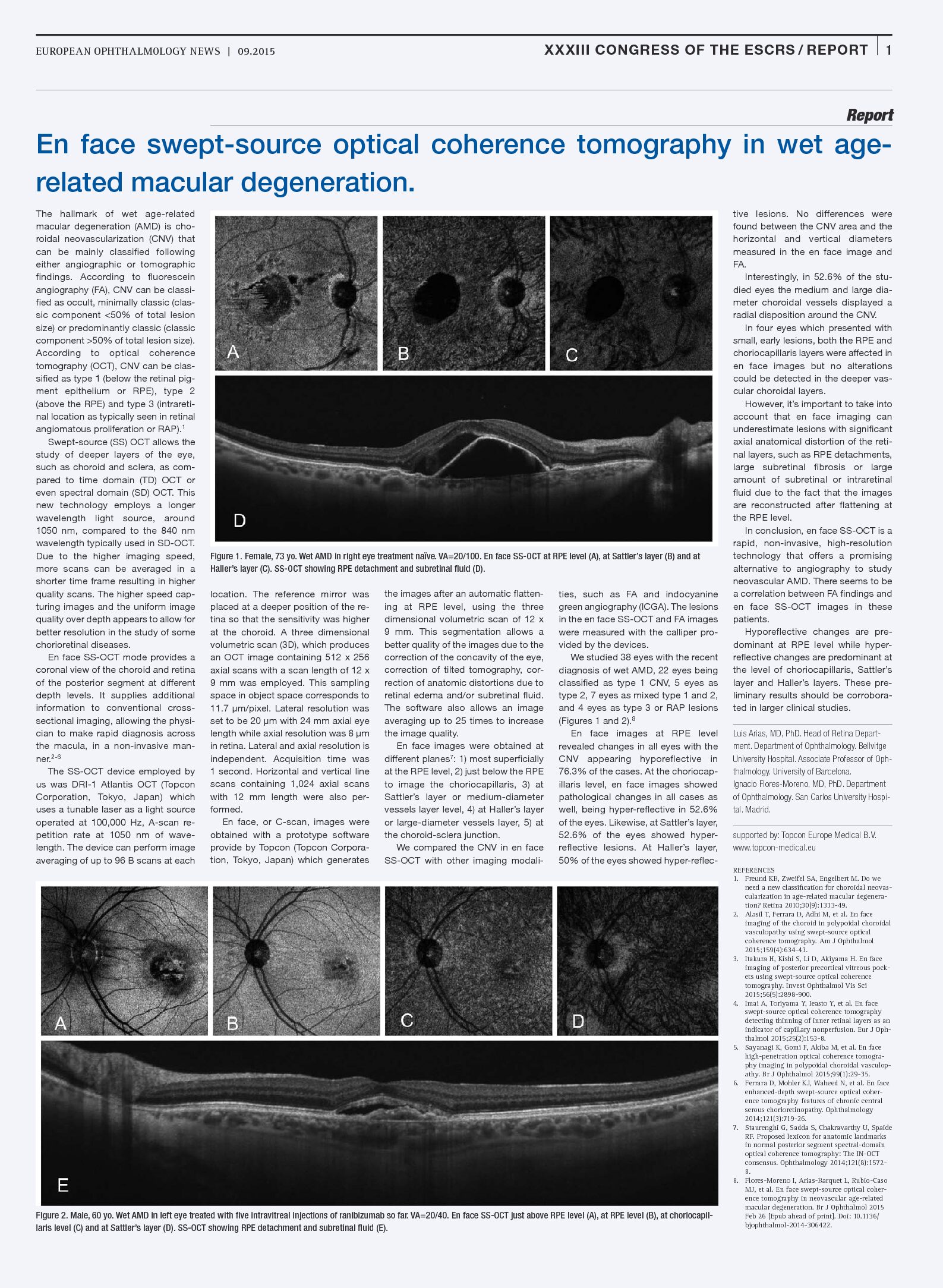
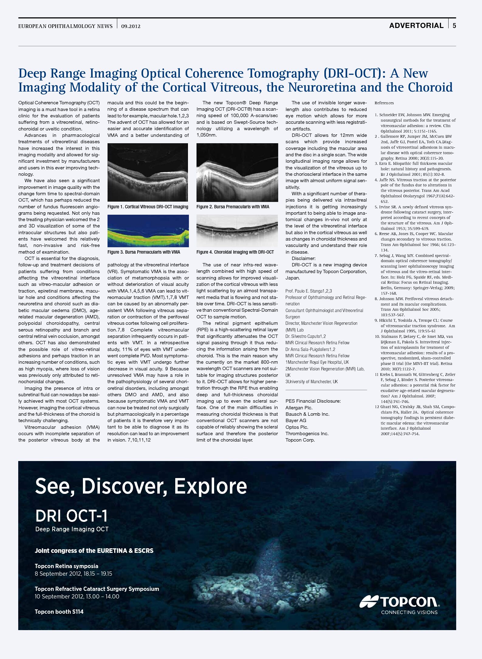
Topcon Healthcare empowers providers with advanced imaging, diagnostic solutions and intelligent data technology, offering a more fully integrated approach to diagnosis and treatment in eyecare.
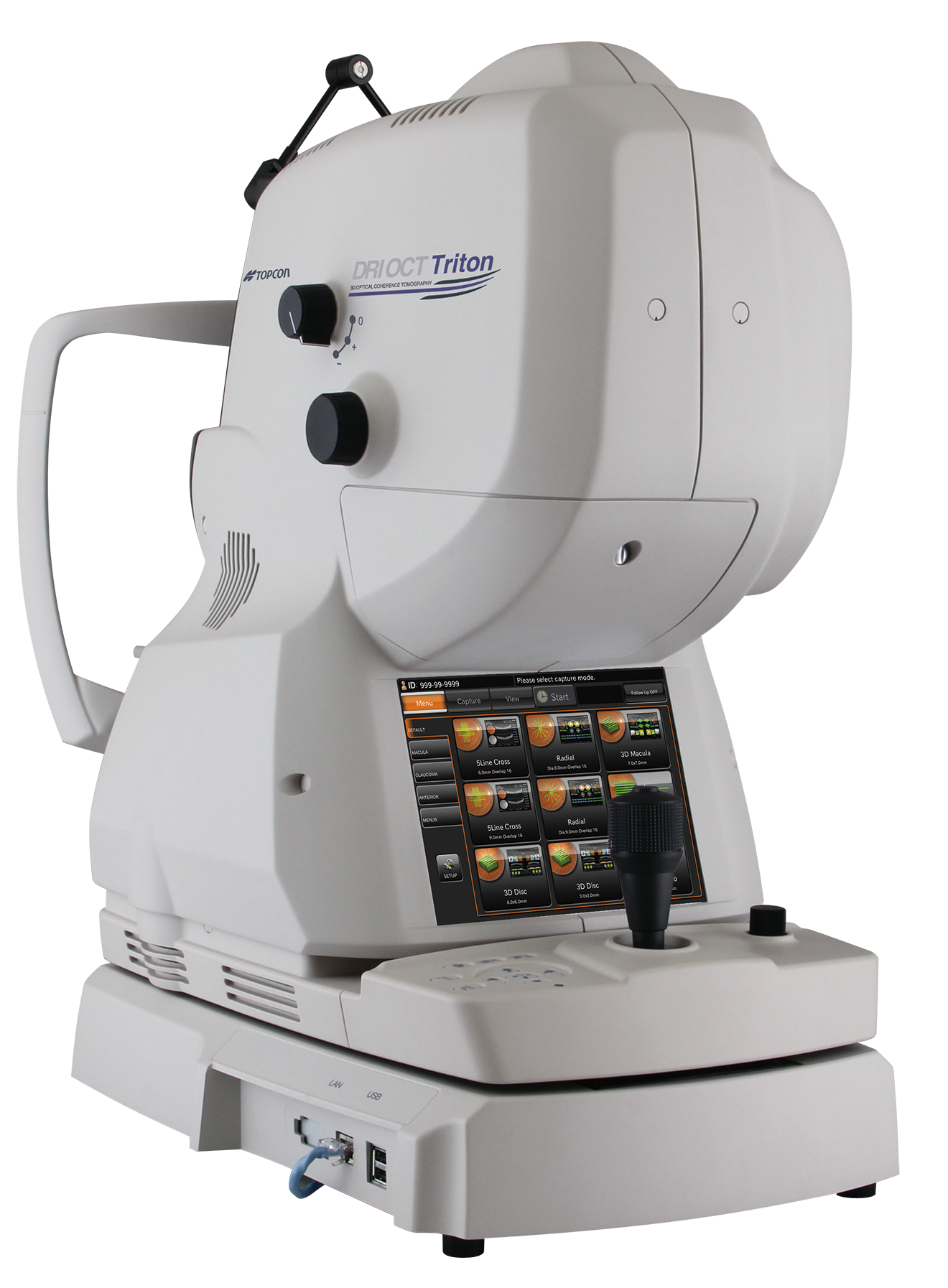
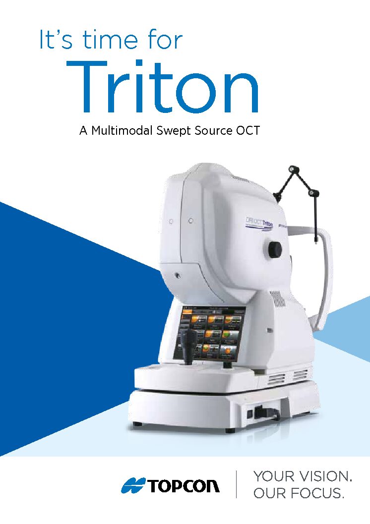
DRI OCT Triton
Swept Source OCT
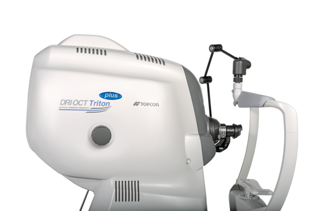
Topcon is the first in the world to introduce a combined anterior & posterior Swept Source OCT, the DRI OCT Triton. The DRI OCT Triton incorporates full color high resolution fundus photography and FA & FAF imaging. FA & FAF imaging is a factory option.
Swept Source technology & 1,050nm wave length
Swept Source OCT provides a significant improvement over conventional OCT. Due to the optimized long wavelength scanning light (1,050nm), there is better penetration of the deeper layers of the eye. Furthermore, this scanning light also penetrates better through cataracts, hemorrhages, blood vessels and sclera.
The world’s fastest scanning speed 100,000 A-Scans/second
Approximately twice higher scan speed, compared to Topcon SD OCT, will bring more scans for a single B-scan image, and more informative image supports efficiency and quality of diagnosis.
Better penetration
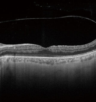
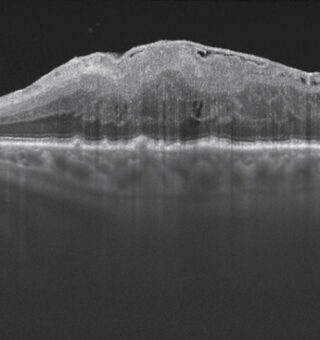
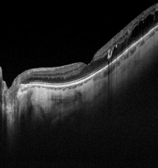
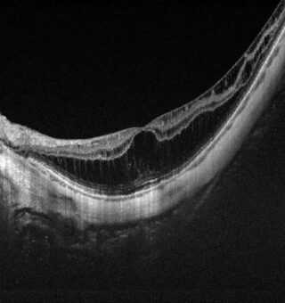
The high penetration of the Swept Source light can easily and clearly visualize deep layers in the eye, such as choroid and sclera. A further benefit of Swept Source is that it can clearly visualize both the vitreous and choroid in a single scan, that are uniformly clear and noise-free. This eliminates the need for time consuming vitreous/choroidal combination scans.
Wide and deep scans
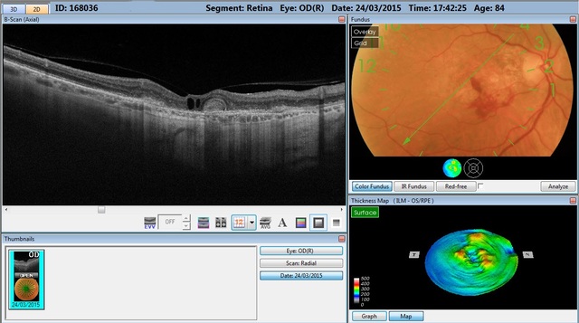
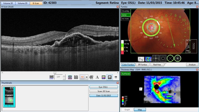
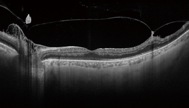
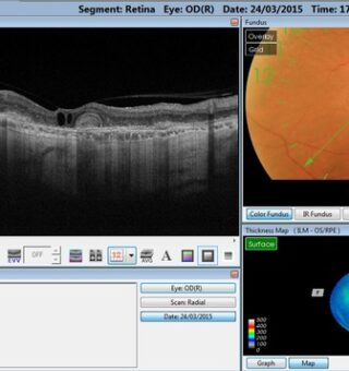
In one single image the vitreous & choroid are revealed in a crystal clear way. The Topcon DRI OCT Triton enhances visualization of outer retinal structures and deep pathologies. The Topcon DRI OCT Triton automatically detects 7 boundaries including the chorio-scleral interface. The 12mm B-scan covers both the macular area and the optic disc.
Invisible scan lines
The invisible 1,050nm wavelength does not distract patients. Patients do not see the scanning line, which is an advantage with elderly patients and children. Reduction in movement artifacts and increased repeatability.
Time efficiency – create one single overview
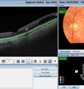
Combination scans cover the macular and disc areas in a single shot, and offer both macular and Retinal Nerve Fiber Layer (RFNL) analysis. Combination scans are time efficient for the operator and convenient for the patient. Combination scans allow both macular and disc analysis in one overview.
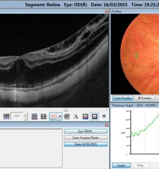
Multi modal fundus imaging
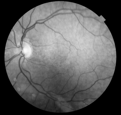
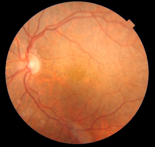
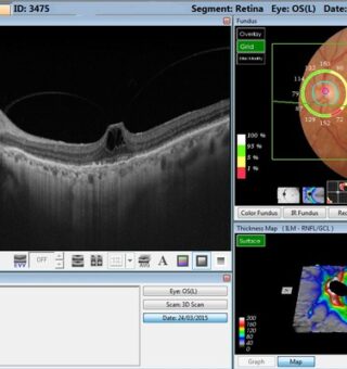
The Topcon DRI OCT Triton offers a true color, non mydriatic fundus image while using a very low intensity flash. This unique feature is a perfect tool for identifying the location of scans in the eye utilizing TOPCON’s patented Pinpoint RegistrationTM. The DRI OCT Triton Plus offers a wide range of diagnostic options with multi-modal color fundus imaging, Fluorescein Angiography (FA) and Fundus Autofluorescence (FAF) for even more diagnostic possibilities. For the first time Pinpoint registrationTM will be available with fundus auto fluorescence and Swept Source OCT.
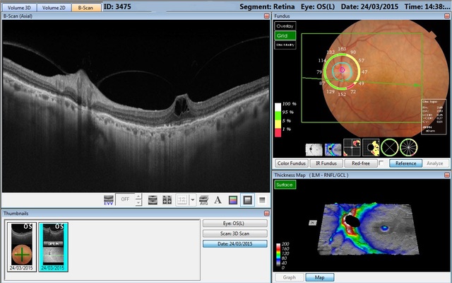
New tracking system – SMARTTrackTM
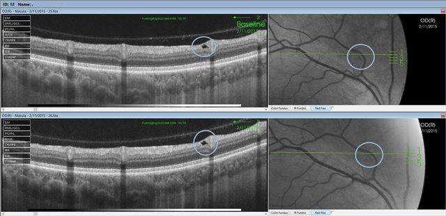
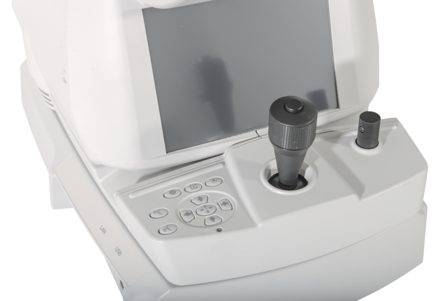
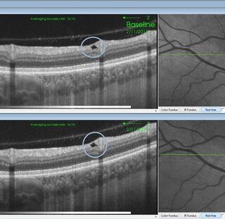
SMARTTrackTM is a very useful tool to compensate for the ever present involuntary eye movements (microsaccades). It allows the automatic acquisition of a follow-up scan in precisely the same anatomical location. SMARTTrackTM enhances the user-friendliness of the machine.
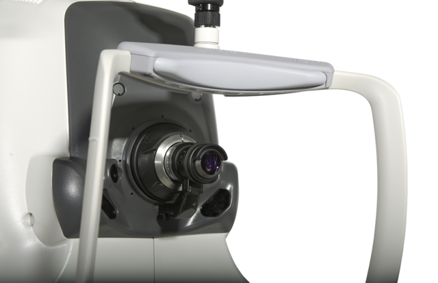
Anterior segment analysis
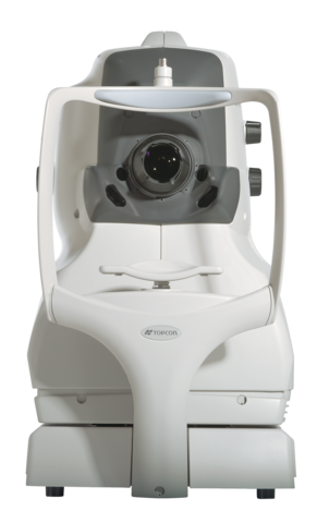
The Topcon DRI OCT Triton can be extended to include anterior imaging, making the Swept Source a versatile diagnosis tool for both anterior and posterior imaging. The anterior attachment ensures sharp images, even in the periphery of the cornea and in depth images of the anterior chamber.
Key Features
- More confident initial diagnosis and ability to track change over time
- Greater clinical efficiency
- Greater patient comfort
- Single scan capture of comprehensive data
| OCT IMAGING: | |
| Methodology | Swept Source OCT |
| Optical light source | Swept Source tunable laser at 1,050nm |
| Scan speed | 100,000 A-scans per second |
| Lateral resolution | 20 µm |
| In-depth resolution | Optical resolution: 8 µm, 2.6 µm digital resolution |
| Photography type | Color, FA*, FAF*, Red-free** |
| Picture angle | 45° |
| Equivalent 30° (digital zoom) | |
| Operating distance | 34.8mm |
| Minimum pupil diameter | ø2.5mm OCT, 3.3mm fundus photo |
| OBSERVATION & PHOTOGRAPHY OF FUNDUS TOMOGRAM: | |
| Scanning range (on fundus) | Horizontal – Within 3 to 12mm |
| Vertical – 3 to 12mm | |
| Scan patterns | 3D scan (12x9mm, 7x7mm, 3x3mm) |
| Linear Scan (Line-scan/Cross-scan/Radial scan) | |
| Fixation target | Internal fixation target |
| Peripheral fixation target | |
| External fixation target | |
| OBSERVATION & PHOTOGRAPHY OF ANTERIOR SEGMENT*** | |
| Photography type | IR |
| Operating distance | 17mm |
| Scan range (on cornea) | Horizontal – Within 3 to 16mm |
| Vertical – Within 3 to 16mm | |
| Scan pattern | 3D scan |
| Linear scan (Line-scan/Radial-scan) | |
| Fixation target | Internal fixation target |
| External fixation target | |
* FA photography and FAF photography can only be performed on the DRI OCT Triton plus
**The color image is processed and is displayed as a pseudo-red-free photographed image
***Observation & photography of anterior segment can be performed only when the anterior segment attachment kit is used.
-
INSERT
-
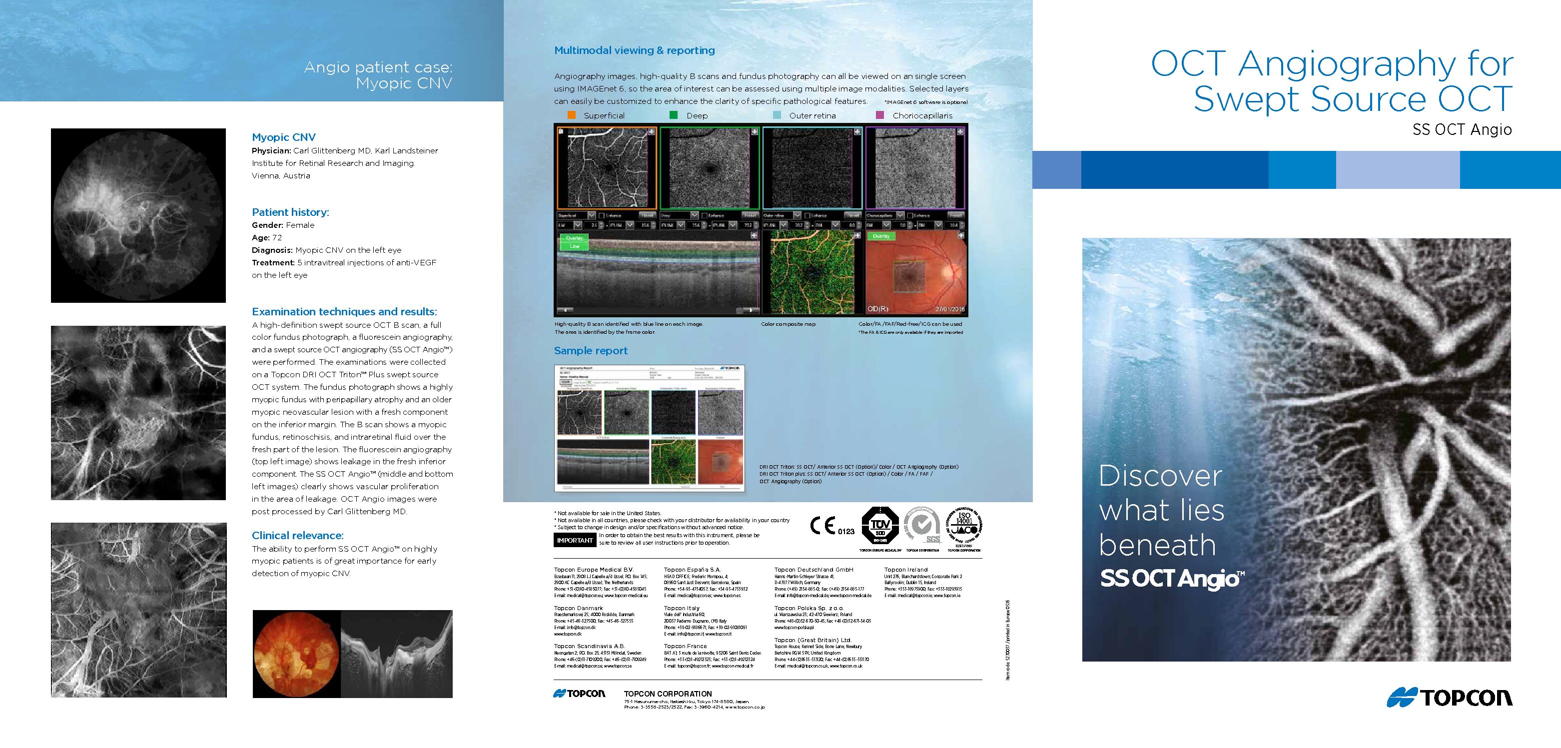
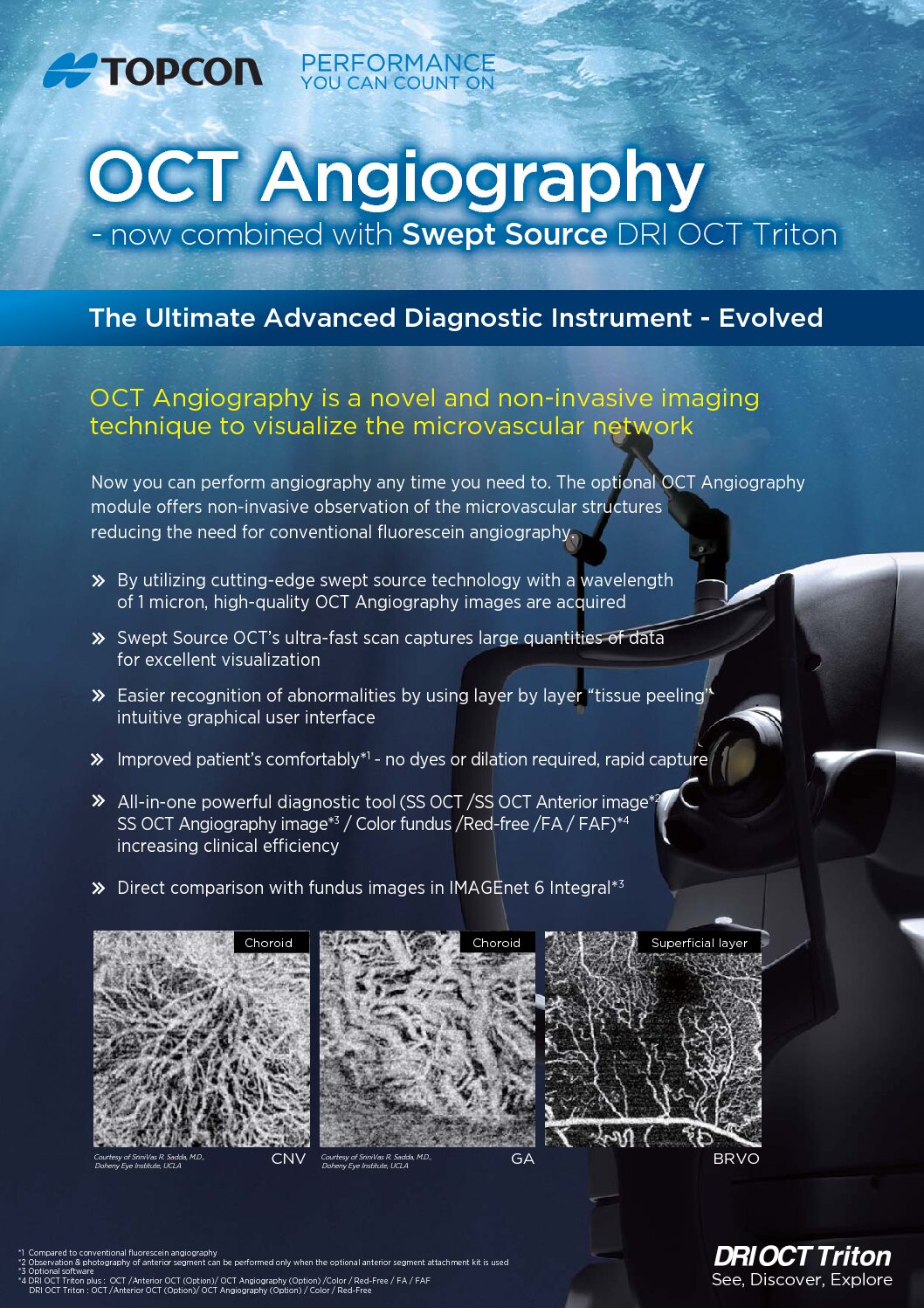
OCT Angiography
-
OCT Booklet
-

Triton Insert
-
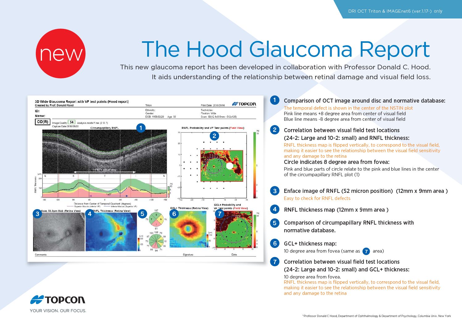
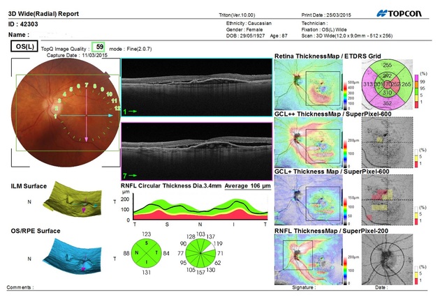
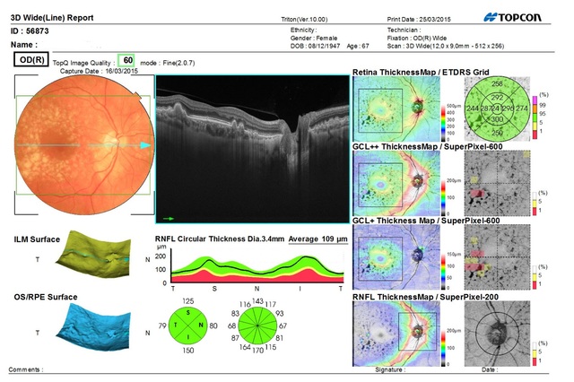
The Hood Report for Glaucoma and Probability Map
-
-
CLINICAL PAPERS
-
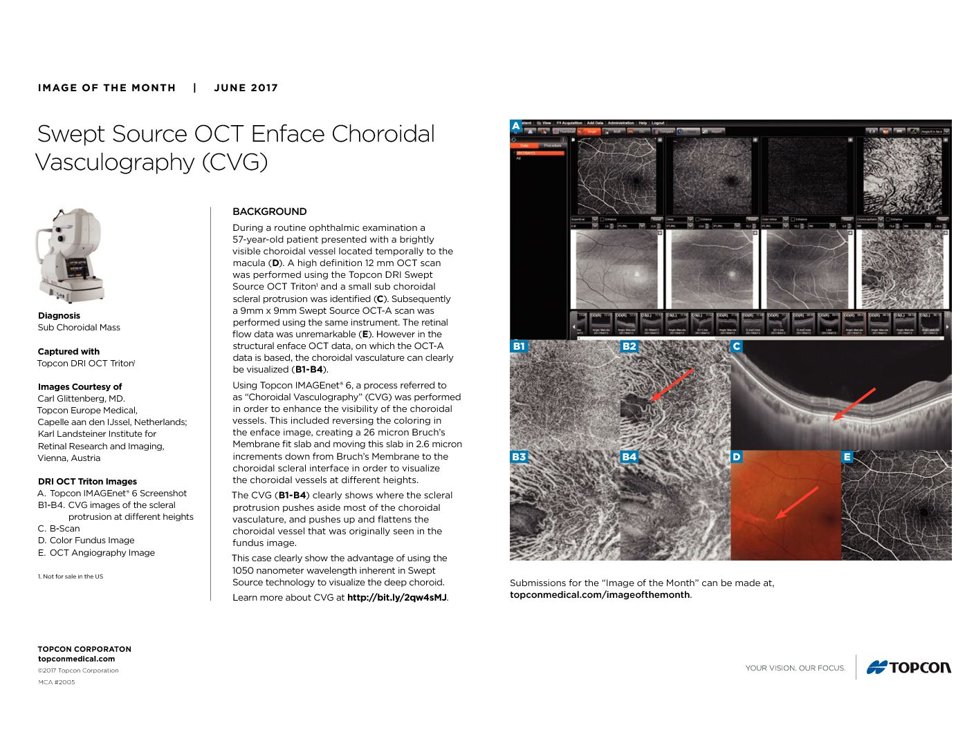
Swept Source OCT Enface Choroidal Vasculography (CVG)
-
Choroidal Imaging with Swept Source OCT
-
EnFace Swept Source OCT
-
Deep Range Imaging Optical Coherence Tomography (DRI-OCT): A New Imaging Modality of the Cortical Vitreous, the Neuroretina and the Choroid
-
OCT Angiography
-
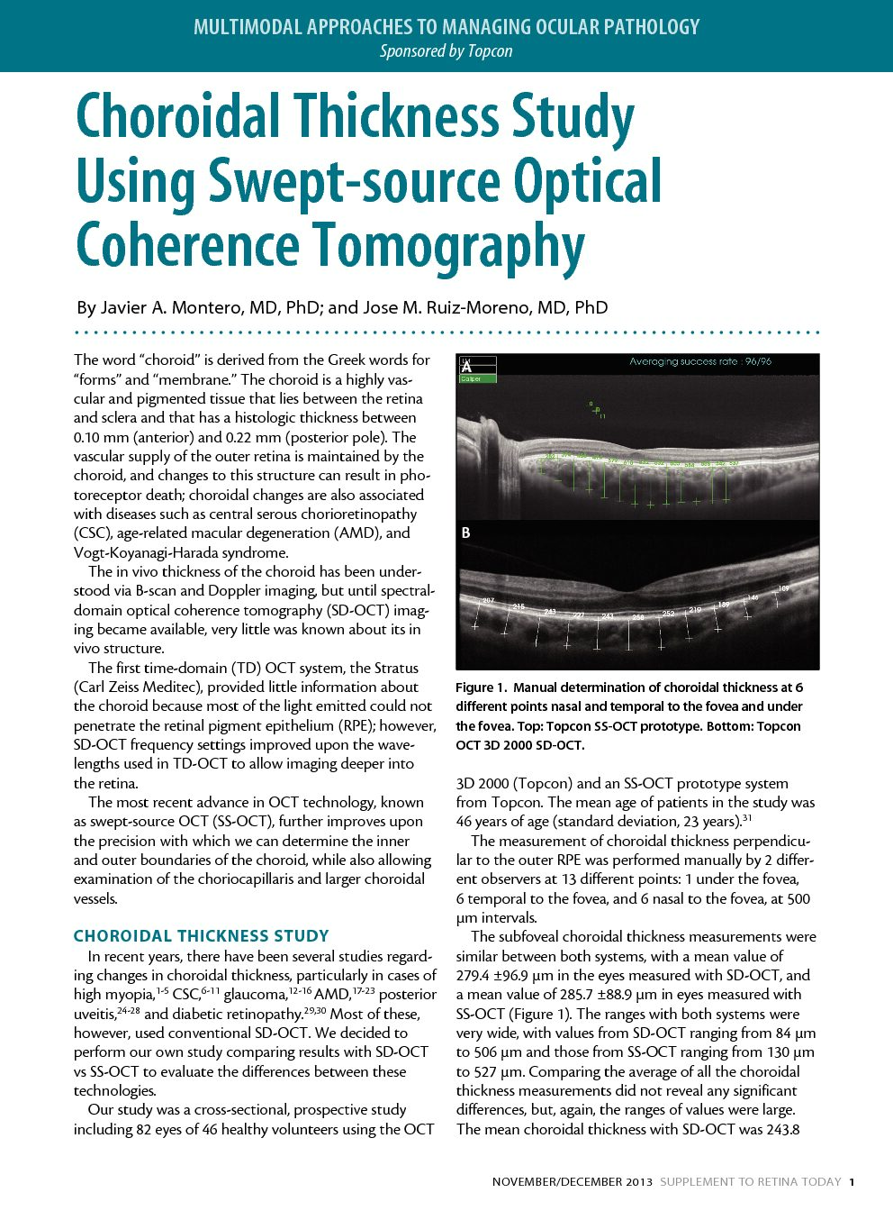
Choroidal Thickness Study Using Swept-source Optical Coherence Tomography
-

Swept-source OCT: Wide-field simultaneous choroid, retina, and vitreous visualization
-
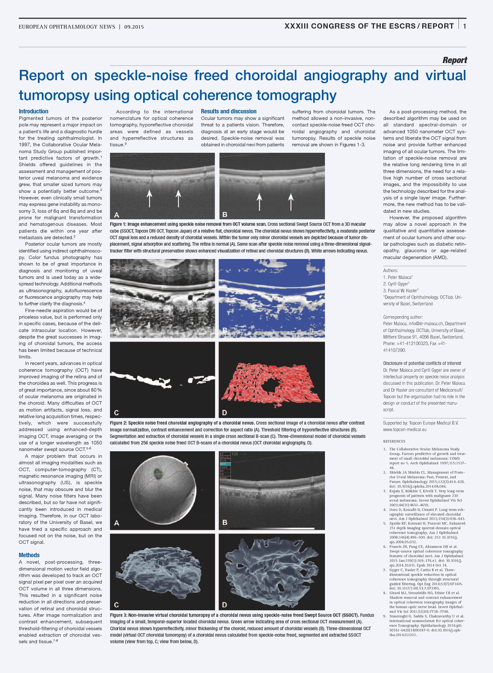
Report on speckle-noise freed choroidal angiography and virtual tumoropsy using optical coherence tomography
-
Swept-Source Optical Coherence Tomography Correlations Between Retina and Choroid Before and After Vitrectomy for Epiretinal Membranes
-
Clinical Advances of Swept-Source OCT and New Non-Damaging Laser Treatments
-
What’s Next in Laser and OCT? Swept-source OCT and Non-damaging Laser Treatment
-
Assessment of Choroidal Topographic Changes by Swept-Source Optical Coherence Tomography After Intravitreal Ranibizumab for Exudative Age-Related Macular Degeneration
-
-
IMAGES
-
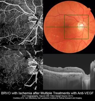
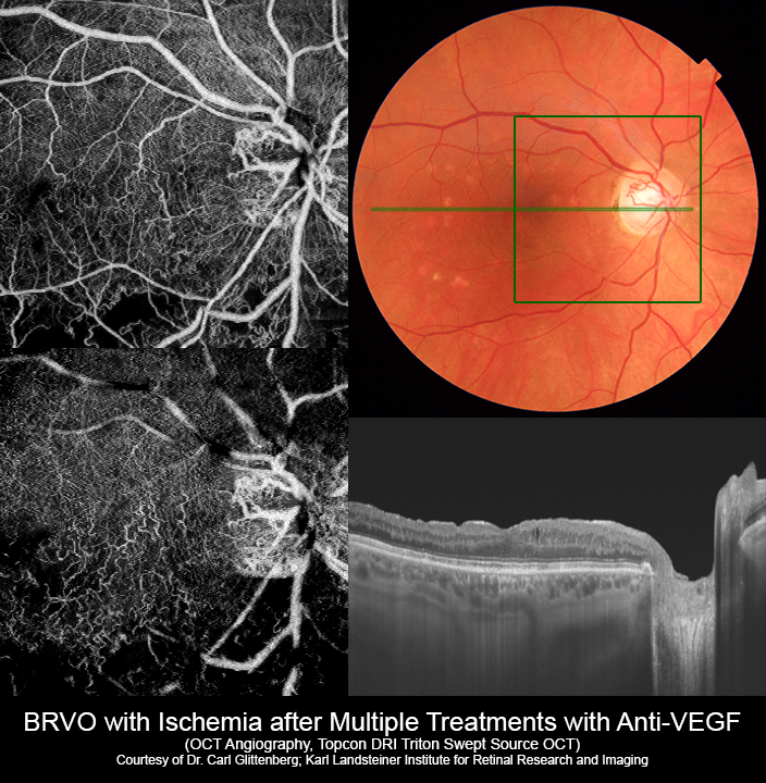
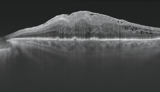
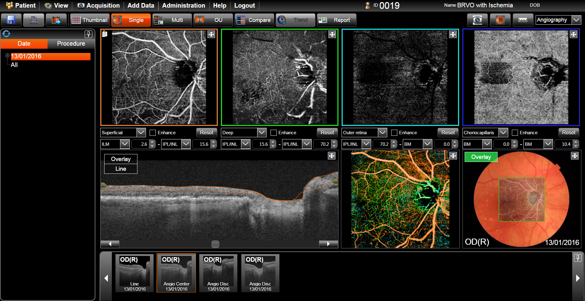
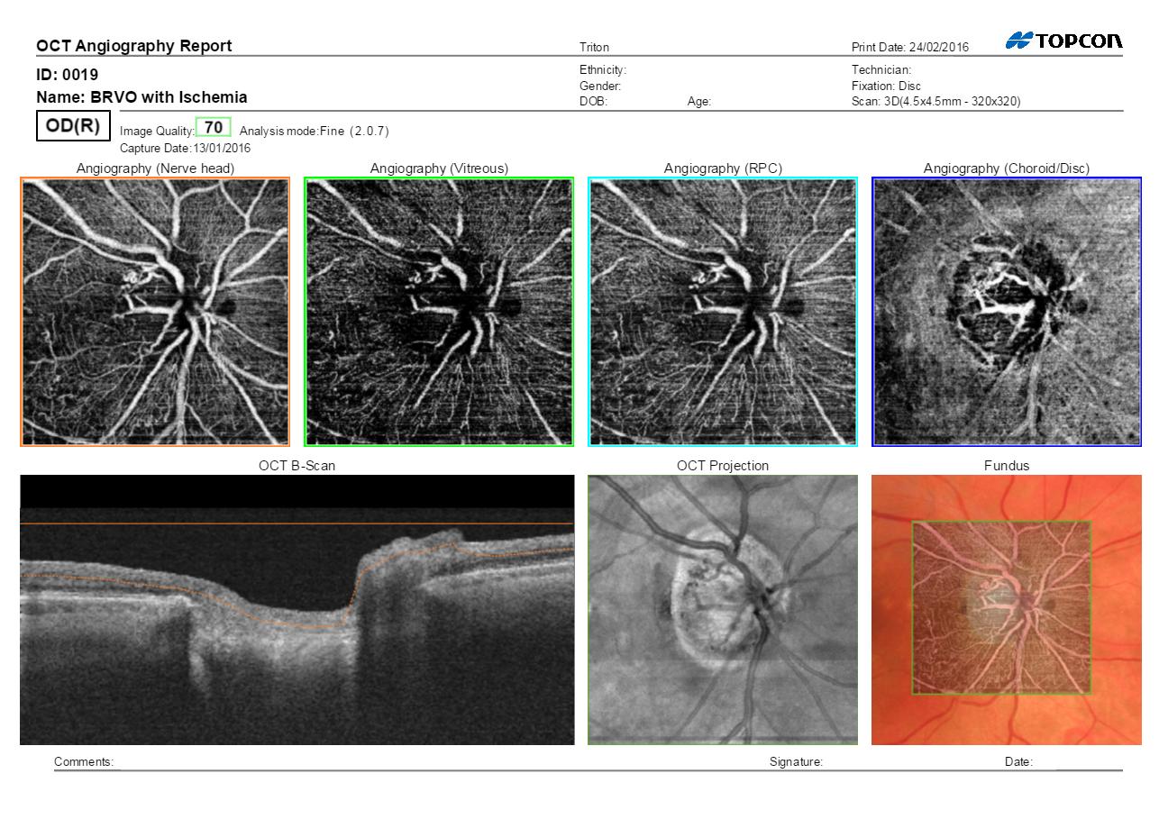
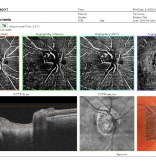
BRVO with Ischemia after Multiple Treatments with Anti-VEGF
-
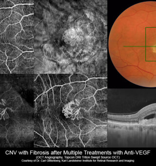
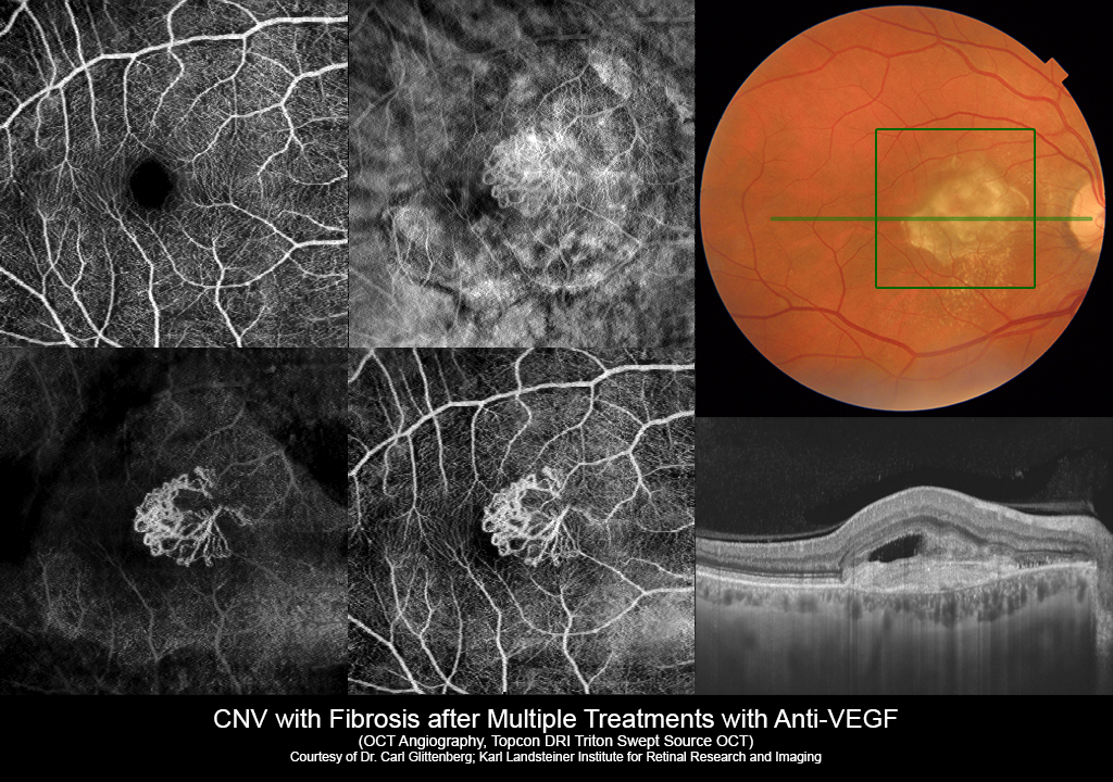
CNV with Fibrosis after Multiple Treatments with Anti-VEGF
-
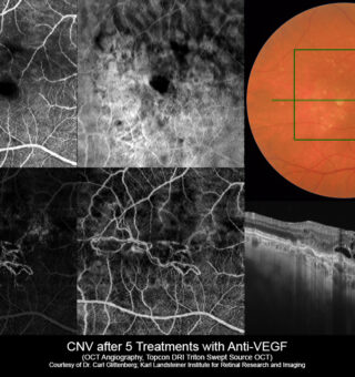
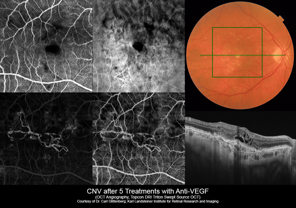
CNV after 5 Treatments with Anti-VEGF
-
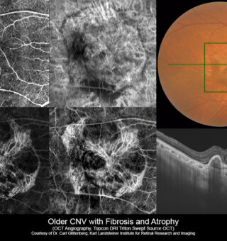
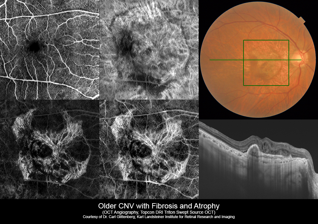
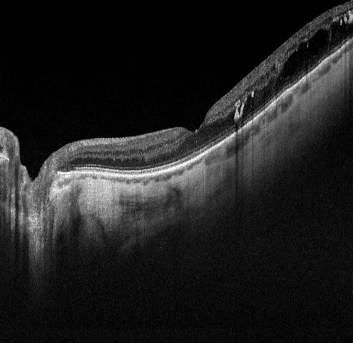
Older CNV with Fibrosis and Atrophy
-
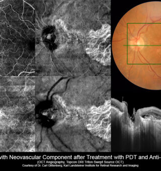
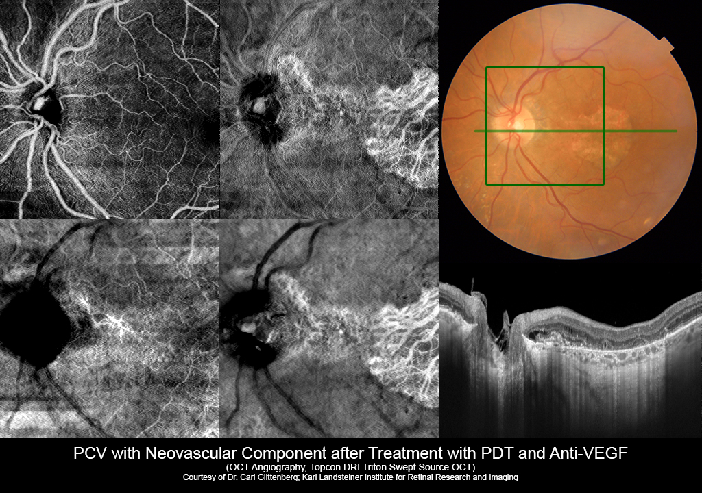
PCV with Neovascular Component aafter Treatment with PDT and Anti-VGEF
-
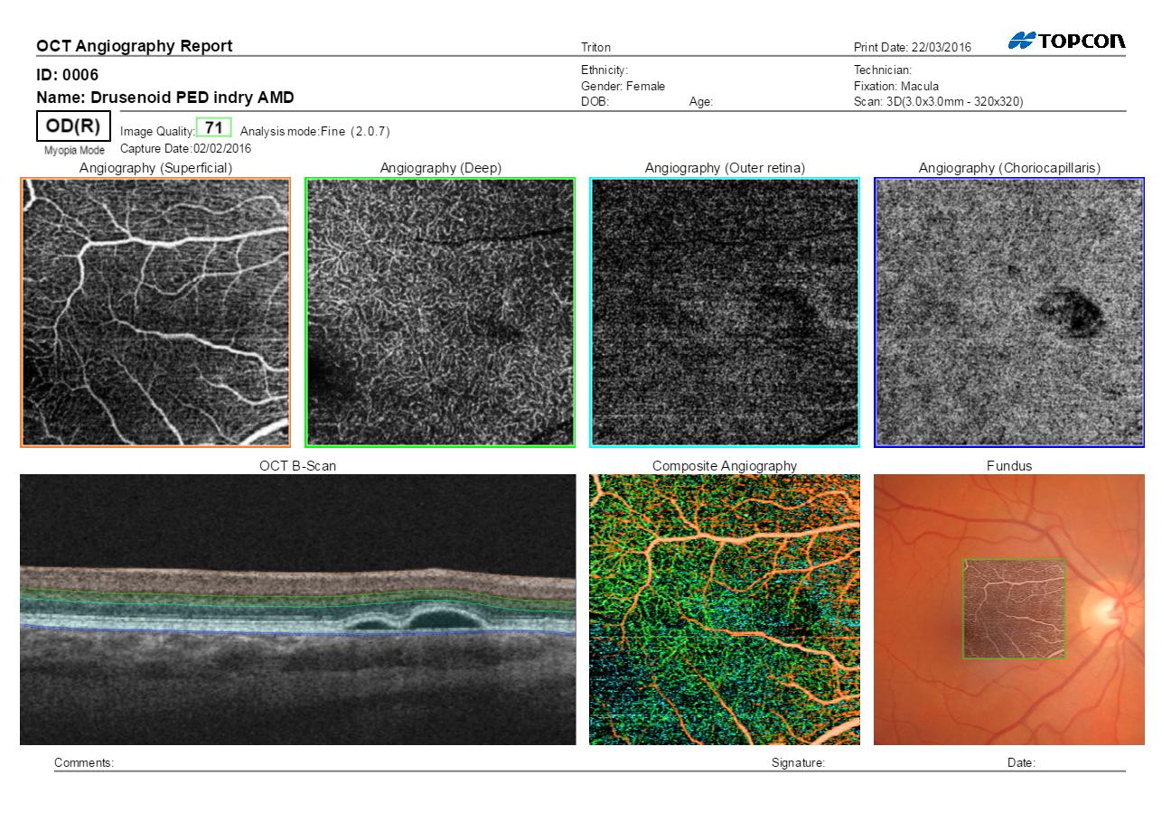
Drusenoid PED Report
-
AMD with Hemorrhages case
-
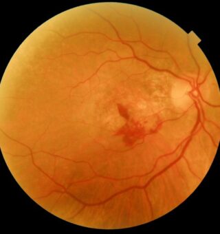
AMD with Hemorrhages case
-
AMD with Hemorrhages Case
-
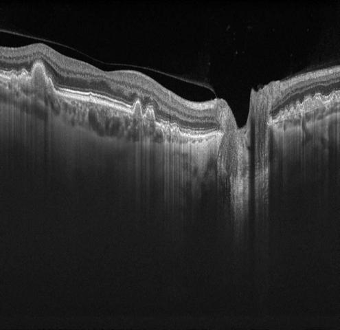
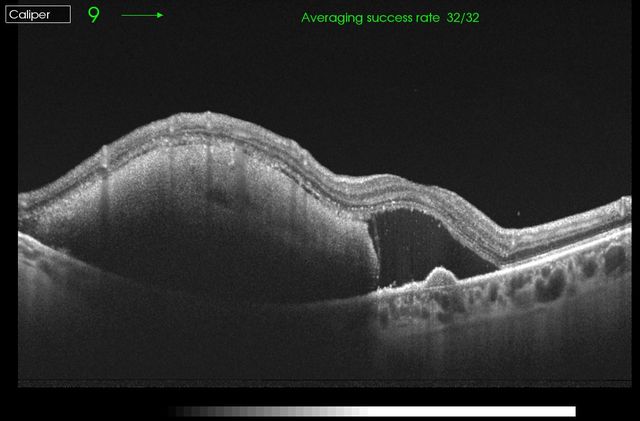
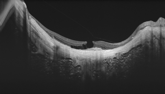
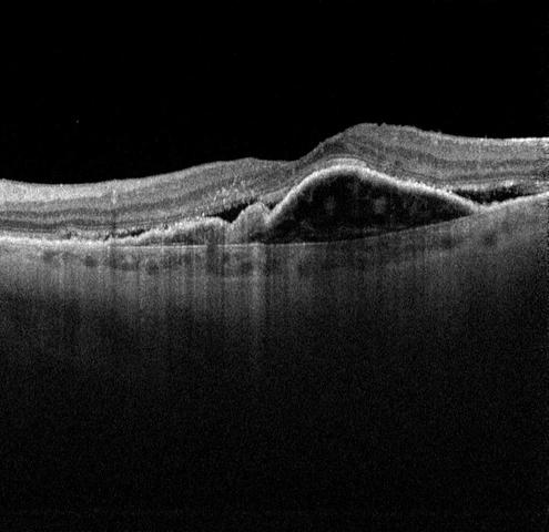
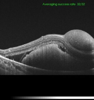
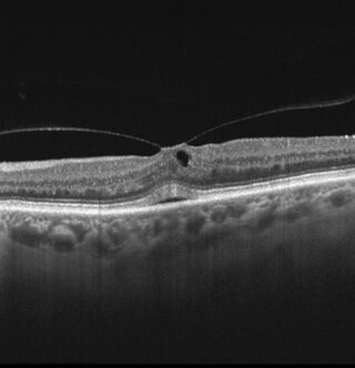
Vitreomacular Traction Case
-
Vitreomacular traction case red-free
-
Vitreomacular traction case
-
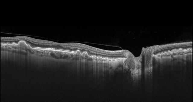
Vitreomacular traction case B-scan
-
Vitreomacular traction case B-scan
-
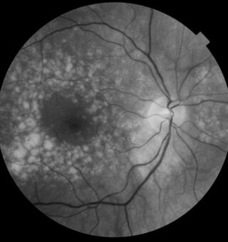
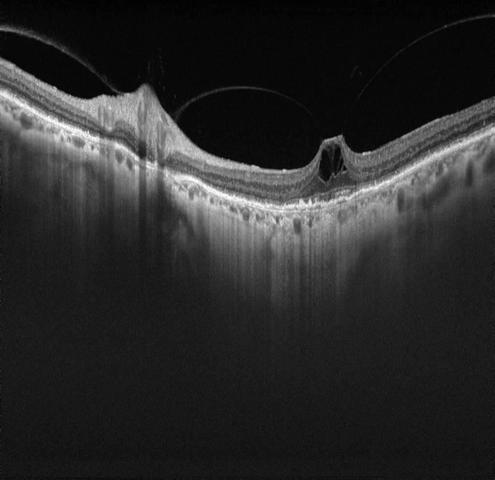
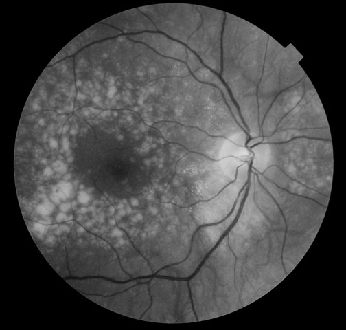
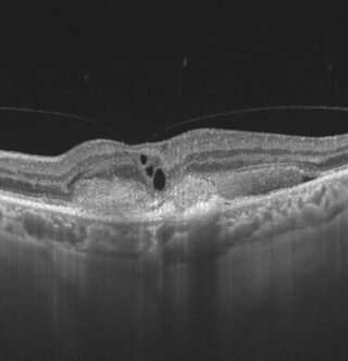
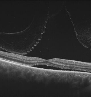
Drusen Case Red-Free
-
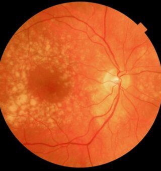
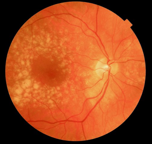
Drusen Case Color
-
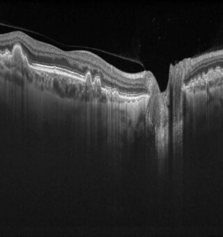
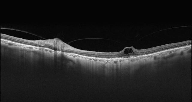
Drusen Case B-Scan
-
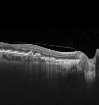
Drusen Case B-Scan
-
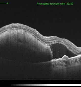
RPED abruption serous with neovascularisation
-
RPED abruption serous with neovascularisation B-scan
-
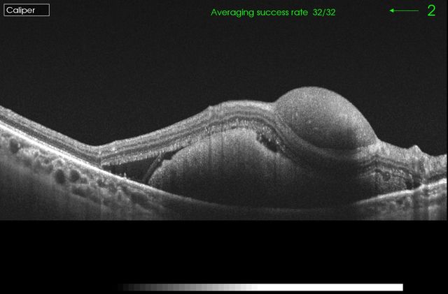
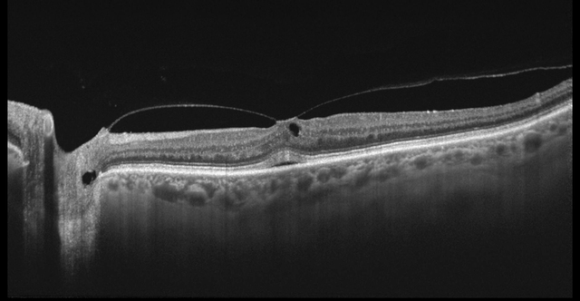
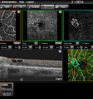
Triton neovascularisation case
-
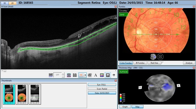
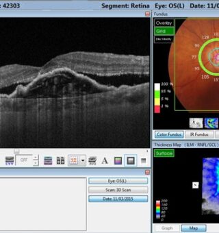
DRI OCT Triton follow up
-
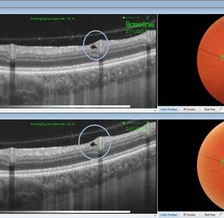
DRI OCT Triton follow up tracking
-
DRI OCT Triton Double Haemorraghe
-
Double Haemorraghe
-
DRI OCT Triton AMD Haemorraghe
-
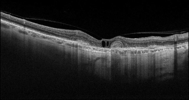
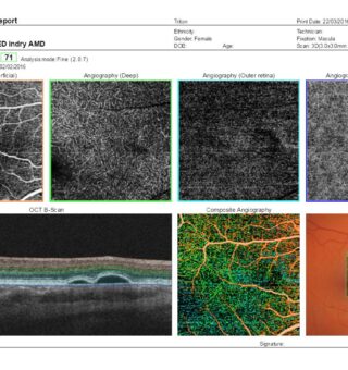
DRI OCT Trition Dry AMD
-
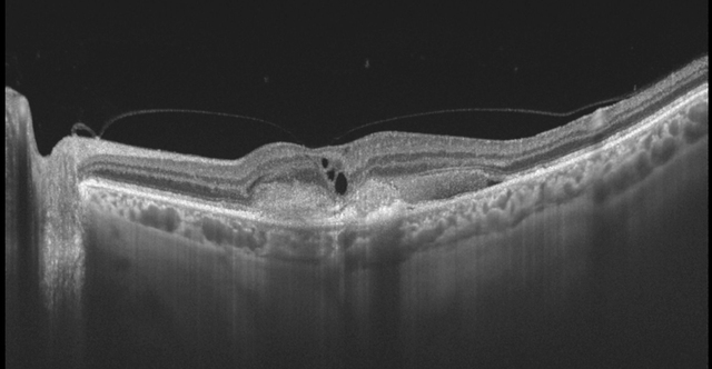
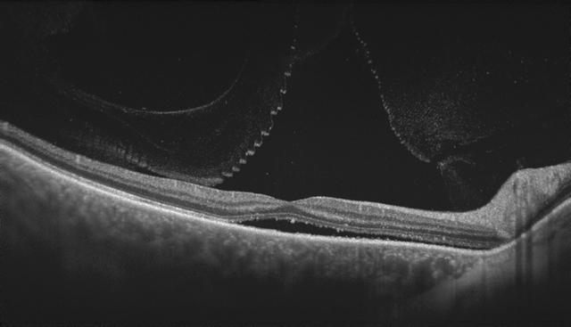
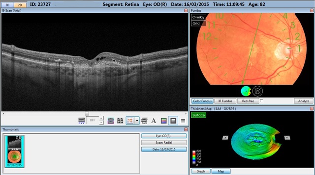
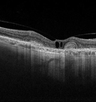
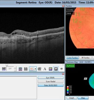
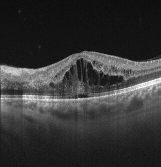
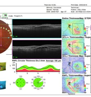
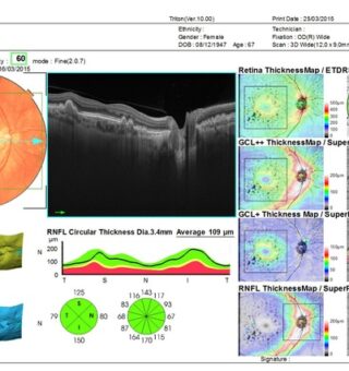
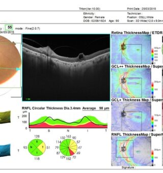
Triton Image
-
Triton Image
-
Triton Image
-
Triton Image
-
Triton Image
-
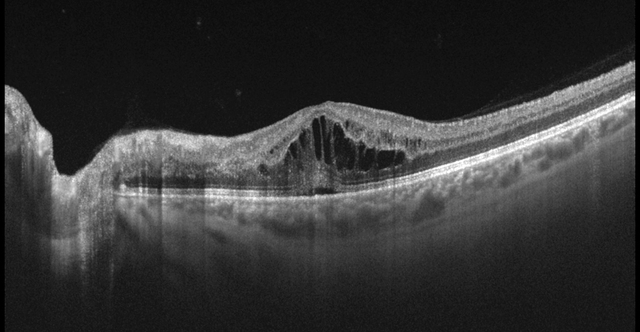
Triton Diabetic retinopathy in myopic eye B scan
-
Triton Diabetic retinopathy in myopic eye -Choroid
-
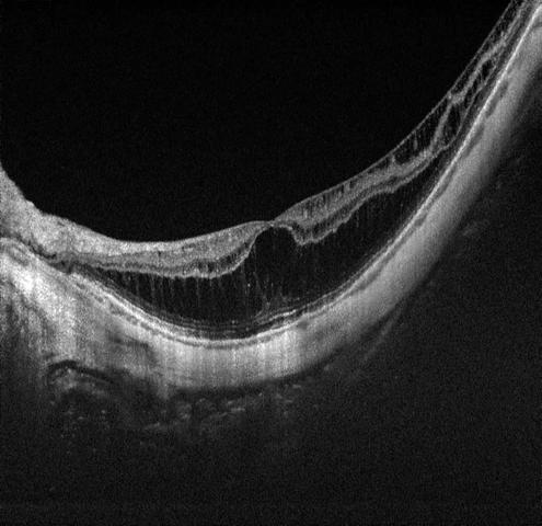
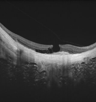
Foveoschisis in highly myopic eyes epiretinal membrane ERM B-scan
-
Triton Foveoschisis in highly myopic eyes epiretinal membrane (ERM)
-

Triton neovascularisation case
-

Triton RPED+ abruption serous with neovascularisation
-
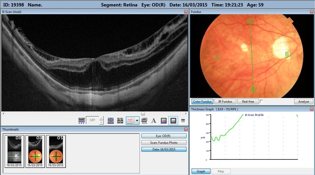
BRVO screenshot
-
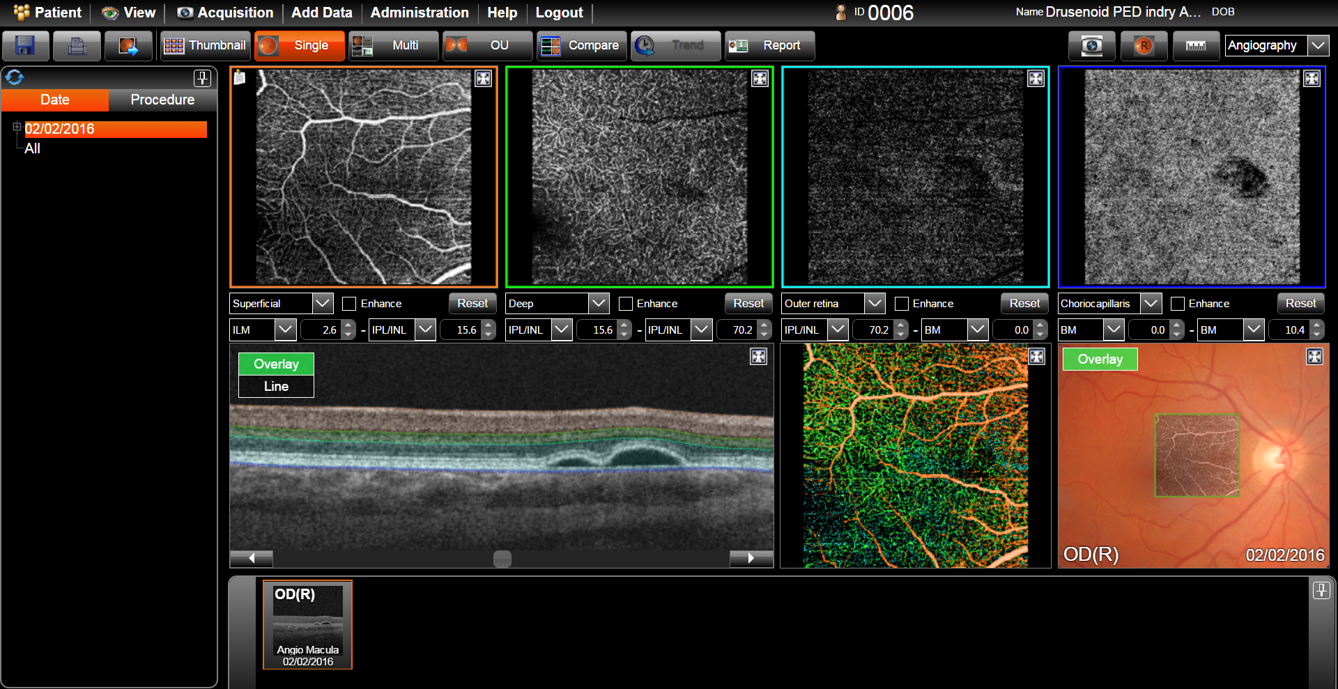
Drusenoid PED screenshot report
-
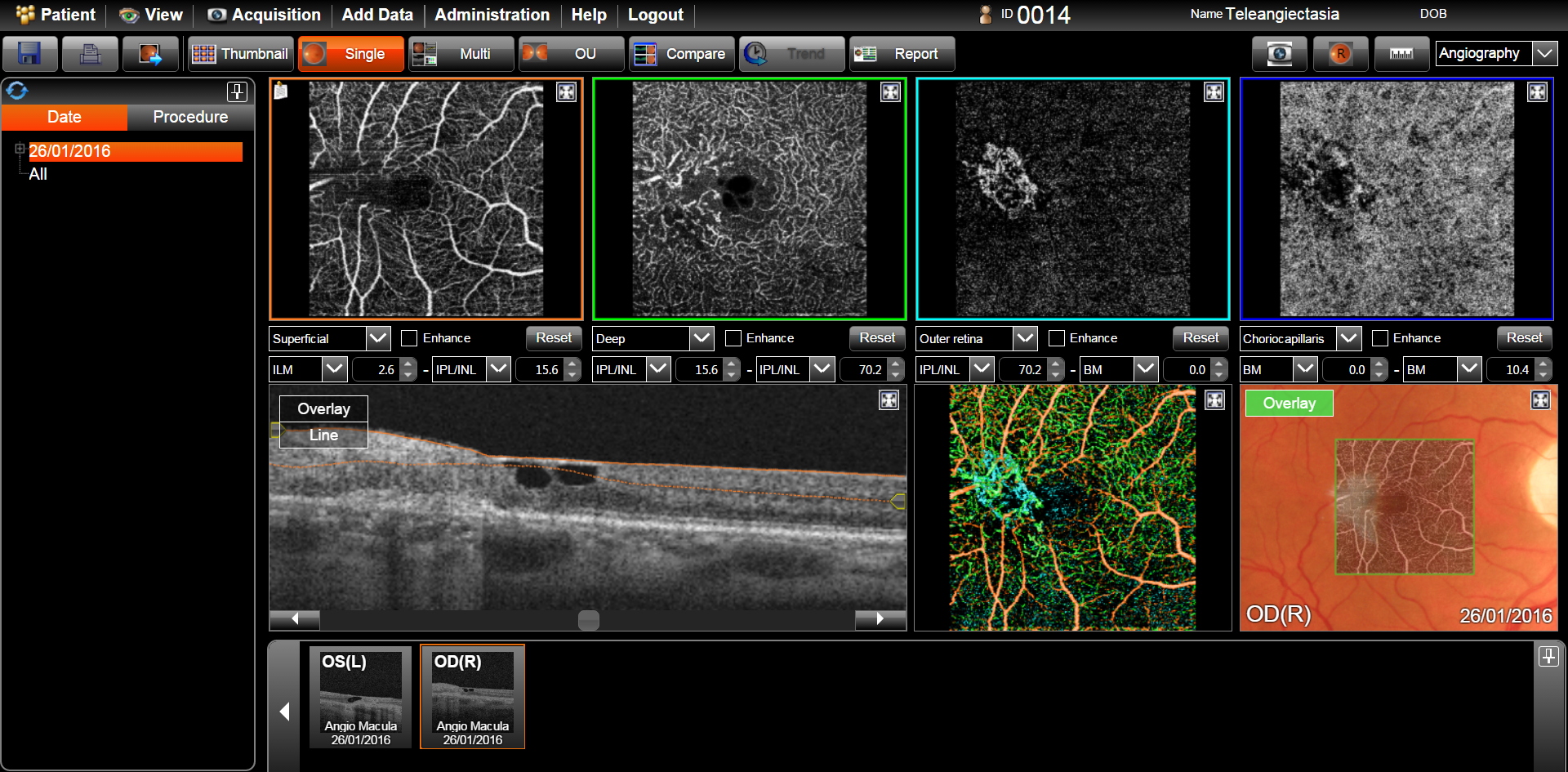
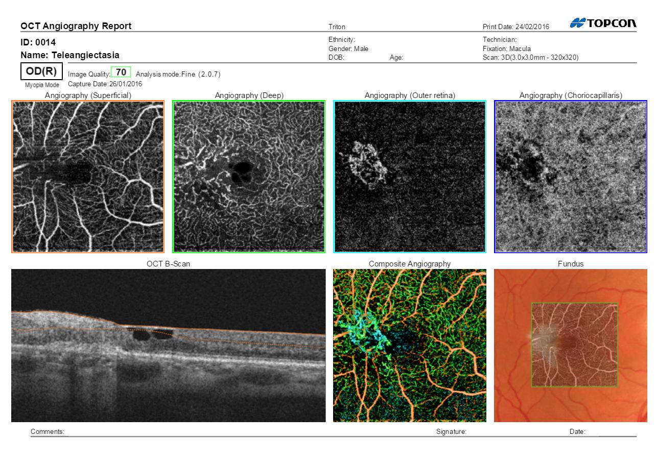
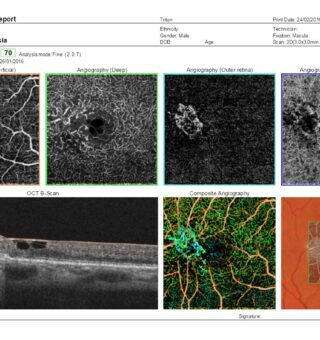
Teleangiectasia screenshot
-
-
SAMPLE REPORT
-
RPED abruption serous with neovascularisation report
-
Soft drusen case report
-
Vitreomacular traction case 1 report
-
Teleangiectasia report
-
Drusenoid PED report
-
BRVO report Angio
-

BRVO report Angio
-
-
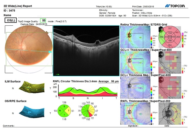
THE HOOD REPORT